Many complex proteins work with actin to produce its many functions. One large family of proteins, called tropomyosins regulates the function of actin filaments in both muscle and non-muscle cells. These proteins consist of rod-shaped coiled molecules.
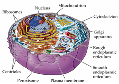
 Actin filaments form a mesh just under the cellular membrane called the cortical network. It links receptor proteins that lie across the membrane to the molecules inside the cell. These
receptors can connect with microbes outside the cell and form a
communication between the outside the cell and the cytoskeleton
structure inside the cell. In this way actin with myosin motors can drag
the virus that is still outside the cell to a better spot for entry.
Actin filaments form a mesh just under the cellular membrane called the cortical network. It links receptor proteins that lie across the membrane to the molecules inside the cell. These
receptors can connect with microbes outside the cell and form a
communication between the outside the cell and the cytoskeleton
structure inside the cell. In this way actin with myosin motors can drag
the virus that is still outside the cell to a better spot for entry.Actin structures are constantly changing as the cell moves and functions—building, breaking down and rebuilding in new ways. Signals from receptors in the membrane direct the changes of actin structures through a series of more than twenty different enzymes called RHO GTPases. These regulate creation of membranes and movement and polarity of cells.
Viruses are particularly focused on manipulating the RHO enzymes. Proteins from viruses alter the actin structures and their functions. It is quite remarkable that viruses can subvert any structure in their way, while they reproduce themselves.
Virus Effects on Actin
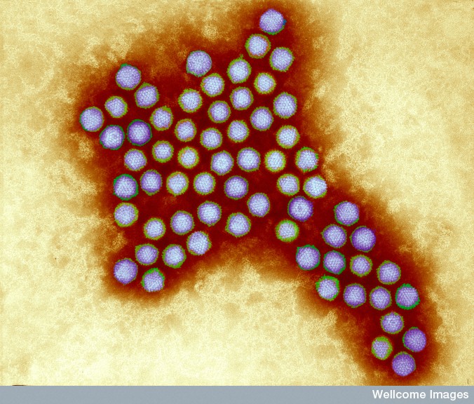 The
first observed effects of adenoviruses on cells were changes in cell
shape, such as becoming more rounded and having more pseudopodia;
although the cells had new pseudopodia, they were unable to move.
Affected cells divided more frequently and piled up on top of each
other. Cells did not have the usual junctures between them. Some of
these changes can occur naturally in a dividing cell, such as the more
rounded shape.
The
first observed effects of adenoviruses on cells were changes in cell
shape, such as becoming more rounded and having more pseudopodia;
although the cells had new pseudopodia, they were unable to move.
Affected cells divided more frequently and piled up on top of each
other. Cells did not have the usual junctures between them. Some of
these changes can occur naturally in a dividing cell, such as the more
rounded shape.Science has finally given us an anatomical place and process in the cell that can respond to Energy and vibration, sound (naturally) but also movement, mind and breath. YAY! we can explain partially why ancient and sacred practices have lasted for generations – they do our cells good.
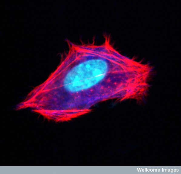
This fabric called the Cytoskeleton likes to be taken out for walks, to stretch, to dance, prance, relax and unwind. Doing yoga, weight lifting, swimming and tai chi all just some of the ways to strengthen and soften the strings of your cells.
Actin
structures are constantly changing as the cell moves and
functions—building, breaking down and rebuilding in new ways. Signals
from receptors in the membrane direct the changes of actin structures
through a series of more than twenty different enzymes called RHO
GTPases. These regulate creation of membranes and movement and polarity
of cells.
Viruses are particularly focused on manipulating the RHO enzymes. Proteins from viruses alter the actin structures and their functions. It is quite remarkable that viruses can subvert any structure in their way, while they reproduce themselves.

 The
first observed effects of adenoviruses on cells were changes in cell
shape, such as becoming more rounded and having more pseudopodia;
although the cells had new pseudopodia, they were unable to move.
Affected cells divided more frequently and piled up on top of each
other. Cells did not have the usual junctures between them. Some of
these changes can occur naturally in a dividing cell, such as the more
rounded shape.
The
first observed effects of adenoviruses on cells were changes in cell
shape, such as becoming more rounded and having more pseudopodia;
although the cells had new pseudopodia, they were unable to move.
Affected cells divided more frequently and piled up on top of each
other. Cells did not have the usual junctures between them. Some of
these changes can occur naturally in a dividing cell, such as the more
rounded shape.
- See more at: http://jonlieffmd.com/blog/virus-tricks-manipulate-the-cytoskeleton#sthash.GSwFgxpZ.dpuf
Viruses are particularly focused on manipulating the RHO enzymes. Proteins from viruses alter the actin structures and their functions. It is quite remarkable that viruses can subvert any structure in their way, while they reproduce themselves.
Virus Effects on Actin
The
first observed effects of adenoviruses on cells were changes in cell
shape, such as becoming more rounded and having more pseudopodia;
although the cells had new pseudopodia, they were unable to move.
Affected cells divided more frequently and piled up on top of each
other. Cells did not have the usual junctures between them. Some of
these changes can occur naturally in a dividing cell, such as the more
rounded shape.- See more at: http://jonlieffmd.com/blog/virus-tricks-manipulate-the-cytoskeleton#sthash.GSwFgxpZ.dpuf